

ESCA Microscopy
ESCA microscopy beamline at Elettra Sincrotrone Trieste
Electron Spectroscopy for Chemical Analysis (ESCA), also called X-ray photoelectron spectroscopy (XPS) is a surface-sensitive quantitative spectroscopic technique that measures the elemental composition in the parts per thousand range. In ESCA microscopy, the X-ray emitted photo electrons are detected and analyzed in terms of their kinetic energy. Small shifts in element specific core or valence energies are produced by changes in surface chemistry and can give detailed information about bond bindings. ESCA is a method sensitive to the chemical information of a few monolayers and therefore offers a special instrument for surface sensitive chemical analysis of heterogeneous materials. The ESCA microscopy beamline at Elettra houses a worldwide unique instrument, the Scanning PhotoElectron Microscope (SPEM), allowing a combination of chemically surface sensitive measurements with high spatial resolution. A beam spot down to 120 nm and energy sensitivity within 180 meV using a third generation X-ray source providing more than 109 photons/s in the probe has opened the opportunity for material science to perform micro-characterization on a spatial scale comparable to that of the processes and phases occurring on morphologically and chemically complex surfaces.
Contact: Luca Gregoratti
Tel: +39 040 375 8025 (office) | +39 040 375 8335 (beamline)
Technical specifications
The 98 periods undulator consists of two equal modules that produce horizontally polarized light. By setting the gap value from a minimum of 13.5 mm up to 40 mm the photon energy can be varied in the range 90÷1500 or 130÷1800 eV when the Elettra storage ring works at 2.0 or 2.4 GeV, respectively. A beam spot down to 120 nm and energy sensitivity within 180 meV, using a third generation X-ray source providing more than 109 photons/s in the probe, has opened the opportunity for material science to perform micro-characterization on a spatial scale comparable to that of the processes and phases occurring on morphologically and chemically complex surfaces.
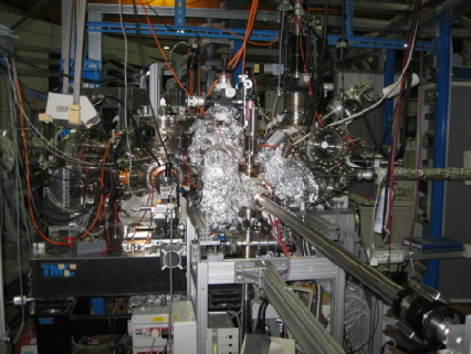
Sample environment
The measurement station consists of three UHV chambers: the experiment chamber, hosting the SPEM, a fast entry lock connected to a small preparation chamber, in which 5 sample holders can be kept in vacuum contemporarily and simple preparation procedures can be performed, and a main preparation chamber with facilities for specimen preparation / characterization. The specimen mounted on a sample holder is transported using magnetic arms and wobble sticks. Additionally, a high pressure chamber can be attached to a cross of the 3 aforementioned chambers.
Detailed information can be found on the beamline’s main homepage.
-
01.02.2024
X-ray Microimaging and Microspectroscopy
-
21.12.2023
AMS-IRMS for radiocarbon dating