

PRESS RELEASE: A multi-modal analysis to characterise fibrotic tissues
|In a recent study an interdisciplinary research team, with scientists operating under the framework of the Central European Research Infrastructure Consortium (CERIC-ERIC), developed and tested a multi-technique approach to characterise the different phenotypes of pulmonary fibrosis (PF).
The team includes researchers from the University of Trieste (Italy), Elettra Synchrotron (Italy), University Medical Center Göttingen (Germany), Max Planck Institute for Multidisciplinary Sciences (Germany) and Georg-August-Universität Göttingen Institute for X-Ray Physics (Germany). The research was published in the scientific journal Computers in Biology and Medicine and represents an important step to the clinical approach of PF, as well as a novel paradigm for tissue analysis.
Pulmonary fibrosis is a disabling disease characterised by progressive involution of the lungs, that gradually impairs the patient’s ability to breathe. It is an irreversible condition that currently has no available curative approaches, and a fatal outcome. Most cases of PF are of the idiopathic type (IPF), a heterogeneous group comprising disorders with very different pathophysiology, and therefore clinical course. It is then essential to improve tissue characterisation techniques – in terms of fiber composition, fiber orientation and micro-mechanical properties- so that each type of IPF can be addressed with the most appropriate treatments, and to develop new ones.
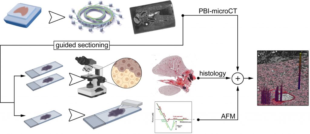
However, structural and functional features of PF cannot be addressed with a single technique; that is why the research team developed and applied a novel multi-technique characterization approach to tissues coming from two different preclinical mouse models: in the first one the disease was chemically induced (by Bleomycin), while in the second one PF was caused by the conditional deletion of an ubiquitin protein ligase (Nedd4-2). Tissue analysis was then based on the combination of three different techniques: classical histopathology, which is hardly conclusive on its own because PF causes heterogeneous and patchy alterations within the lungs which are similar in their appearance in all different PF types; propagation-based phase-contrast micro computed tomography (PBI-microCT), used to assess structural alteration (fibrotic loci) in whole fixed and paraffin embedded lungs, thus facilitating novel targeted microtome sectioning of planes of interest; atomic force microscopy (AFM), useful to assess changes in the local tissue mechanical properties, especially stiffness, of the previously identified altered structures.
Through the combined application of these complementary investigative techniques, the researchers were able to discriminate samples from the Bleomycin-induced model from those of genetically altered mice (Nedd4-2), thus distinguishing the different pathophysiology leading to IPF.
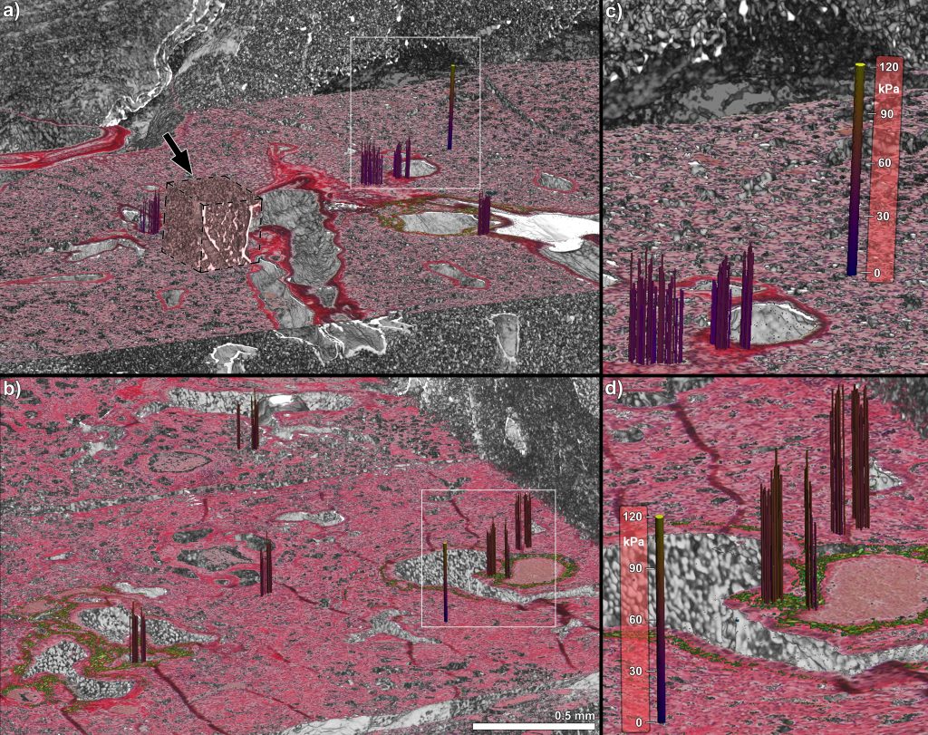
Spatial correlation at the near-cellular level of phase contrast micro tomography, histology and atomic force microscopy
However, more than a point of arrival, this new pipeline of investigation could represent a new approach to the analysis of tissue samples: “Formalin Fixed Paraffin Embedded (FFPE) tissues are the most common way used worldwide in the hospitals to preserve vast and valuable patient material. Therefore, the access to a tissues characterization by means of more techniques, rather just histology, could lead to deeper investigation of pathologies that are still not understood completely “– explains Lorenzo D’Amico, CERIC PhD fellow which carries out his research at the Synchrotron Radiation for Medical Physics (SYRMEP) beamline at Elettra Synchrotron, and first author of the paper.
CERIC-ERIC is a research infrastructures consortium founded by the European Commission in 2014. It offers researchers and industry a single point of access to more than 60 techniques and laboratories in eight Central and Eastern European countries for multidisciplinary research at the micro- and nano-level in the fields of advanced materials, biomaterials and nanotechnology.
ORIGINAL ARTICLE:
PRESS FOLDER: https://drive.ceric-eric.eu/d/afd5813047334d4b8b26/
- PDF of the press release;
- Video: spatial correlation at the near-cellular level of phase contrast micro tomography, histology and atomic force microscopy;
- Fig 1 – Graphic description of the research work and of the complementary analyses performed;
- Fig 2 – 3-dimensional integration of phase contrast micro tomography, histology and atomic force microscopy.
CONTACTS: CERIC-ERIC Press Office: press@ceric-eric.eu
Marcello Turconi, marcello.turconi@ceric-eric.eu
-
12.04.2024
Call for Executive Director
-
12.04.2024 | call for proposals, Research
20th Call for Proposals: an overview